


The pain of endometriosis – making a diagnosis (Part Two)
In the previous installment of this essay, we explored some of the reasons for the delay in the diagnosis of endometriosis and diagnostic tools used.
Like all medical conditions, the diagnosis of endometriosis follows the well-established routine of history taking, physical diagnosis, and ordering targeted tests that can either rule in, rule out or confirm the diagnosis of the condition.
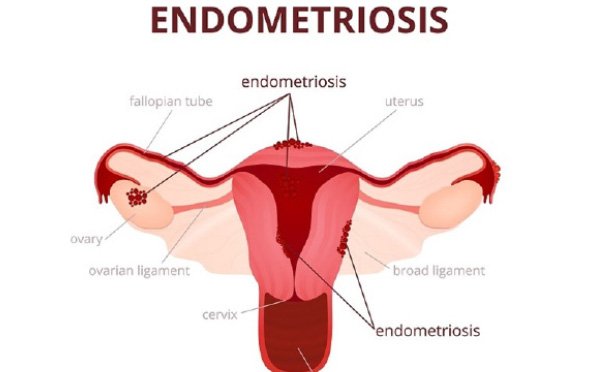
One key element of its nature is pain, wherever it may occur, coming during the menses. The pain of endometriosis may come a week or two weeks before the onset of the menses, become severe during the menses and wane after the period, only to repeat its course in subsequent cycles. The key here is the cyclicity of the pain with respect to the menses. As mentioned in the previous instalment, the severity of the pain has little to do with the stage (severity) of the disease. This week continues the discussion on the different diagnostic tools which included Vaginal scan, Endometriosis mapping and Barium Enema(Sepeiti).
COLONOSCOPY
Colonoscopy involves passing a camera into the large bowel up to the junction of the large and small bowel. This test can also be done in cases of bowel related symptoms of endometriosis including bleeding during bowel motions. Its advantage over a barium enema is its ability to take a biopsy (sample) of the lesion (diseased area) to confirm a diagnosis before the operation can be done.
MRI SCAN
Advanced testing with an MRI scan is possible in selected cases. An MRI scan is capable of detecting smaller endometriotic nodules in the bowel, bladder and uterine ligaments. It is great at picking endometriosis of the womb (adenomyosis). Like other investigations it can only pick large areas of fibrosis occasioned by endometriosis. A negative result does not rule out minimal and mild disease. The down side of this investigation is the considerable cost to the patient.
MRI AND EXAMINATION
UNDER ANAESTHESIA
Another important investigation is examination under anaesthesia. This involves doing a digital vaginal and rectal examination while the patient is sleeping under the influence of anaesthetic agents. The benefit of examination under anaesthesia is that the patient’s vaginal and pelvic muscles would be relaxed allowing thorough exploration of the vaginal wall, the pelvis, the bowel, uterine ligaments, assessment of the mobility of the uterus and ovaries. It also gives detailed information as to whether the window behind the uterus has been obliterated by this destructive disease.
A study comparing the ability of different tests to pick up endometriosis when present, comparing ordinary vaginal scan, a rectal scan, sigmoidoscopy (camera up the rectum and stopping only in the sigmoid colon), MRI and examination under anaesthesia found that the best tool for diagnosis was the use of the old digital vaginal examination under anaesthesia. The down side to this mode of investigation is that one has to incur theatre fees and a day hospital bed fee, making it more expensive.
It is possible however, that digital examination under anaesthesia may be offered in the outpatient setting in clinics with a procedure room and availability of an anaesthetist or nurse anaesthetist. Only in this setup is digital examination under anaesthesia cost effective. It is noteworthy that the normal digital examination without anaesthesia has its uses, but falls short of defining the extent of the spread of endometriosis in the pelvis thereby limiting holistic planning for the operation where different specialists may be required to co-operate on the management of the patient.
DIAGNOSTIC LAPAROSCOPY
The definitive test for endometriosis remains diagnostic laparoscopy. This is an operation through a key-hole in which the patient is not opened in the usual way. At laparoscopy 2-5 holes, wide enough to fit a pen are made on the tummy. The gynaecologist then gains access into the abdominal and pelvic cavities through these holes. A tiny camera is then inserted at the belly button which then becomes the eye of the surgeon through which he would then search for endometriosis in the pelvic cavity.
A biopsy of the endometriotic lesions would be required to make a definite diagnosis. Without a biopsy, diagnostic laparoscopy over-diagnose endometriosis in up to 50% of patients, leading to unnecessary treatment for a condition that is none existent. A diagnostic laparoscopy is valuable in defining the extent of the disease, the organs affected, and planning for the next stage in the treatment phase. It facilitates referral to a specialist surgeon and informs the kind of team that need to be assembled to tackle the menace that is endometriosis.
It however pales in significance in terms of cost savings, when compared to endometriosis mapping scan. As seen earlier, endometriosis mapping allows for a diagnosis of severe deep infiltrating endometriosis, defines the extent of disease, defines the team mixture of surgeons required to tackle the disease from the outset, cutting unnecessary diagnostic laparoscopic surgery. In the diagnosis of endometriosis, diagnostic laparoscopy has its place in non-deep infiltrating endometriosis which is the domain mostly of minimal, mild to moderate disease.
*In the next instalment, we will discuss treatment options for endometriosis and evaluate their effectiveness.
Dr Vincent G Molelekwa is Obstetrician, Gynaecologist, Fertility Specialist, Endoscopic Surgeon, Gaborone Fertility Clinic